
Figure 4. Effect of cortical inactivation on the localization of 40 ms noise bursts.
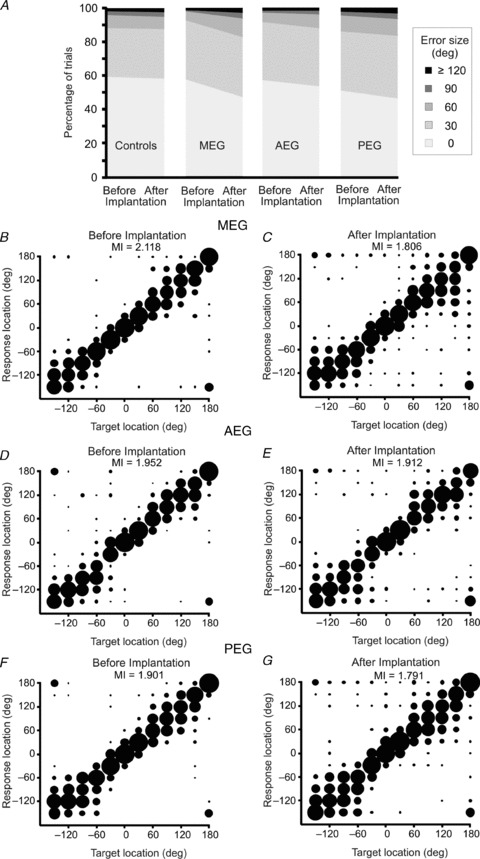
A, distribution of trials classified by error size before and after bilateral implantation of either drug-free Elvax (Controls) or muscimol-Elvax over the cortical regions indicated (MEG, AEG or PEG). B–G, stimulus–response plots showing the approach-to-target performance before (left panels) and after (right panels) implanting muscimol-Elvax bilaterally over the MEG (B and C), AEG (D and E) or PEG (F and G). The MI (in bits) between target location and response location is given in each panel. Stimulus–response plots for the control group are not shown as there were no differences following Elvax implantation (MI = 1.921 bits before implantation and 1.973 bits after implantation).