
Figure 8. Muscimol-Elvax implants do not modify the thickness of cortical layers.
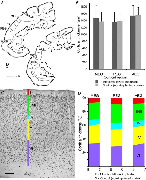
A, drawings of coronal sections of the left hemisphere at two antero-posterior levels of the EG showing where the cortical thickness was measured in the three different auditory regions. Scale bar, 2 mm. B, mean ± SD cortical thickness in the region where muscimol-Elvax was implanted and in a corresponding region of non-implanted auditory cortex from the same brain. C, photomicrograph of a coronal section showing NeuN immunoreactivity to reveal the different cortical layers in a region of the PEG where muscimol-Elvax had been implanted. Scale bar, 200 μm. D, relative thickness of each layer in the region where muscimol-Elvax was implanted and in a corresponding area of non-implanted auditory cortex from the same brain. AEG, anterior ectosylvian gyrus; D, dorsal; LV, lateral ventricle; M, medial; MEG, middle ectosylvian gyrus; PEG, posterior ectosylvian gyrus; pss, pseudosylvian sulcus; sss, suprasylvian sulcus.