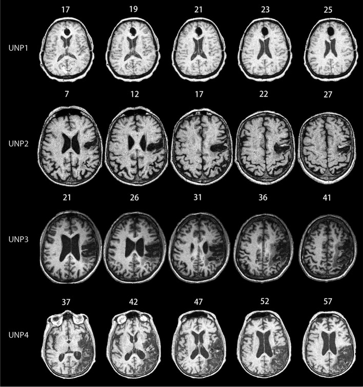
Figure 1.
MRI scans for all of the unilateral neglect patients (UNPs) included in the study. Numbers listed above each picture depict the MNI coordinates. UNP1 has lesions in the anterior cingulate, orbitofrontal, and thalamic regions. UNP2 has lesions in the frontal, parietal, and occipital regions. UNP3 has lesions in the posterior frontal and anterior parietal regions. UNP4 has lesions in the parietal regions and temporal regions, extending into the temporal-occipital junctions. We include a figure depicting slice regions for each participant in the Appendix, Figure A1.

