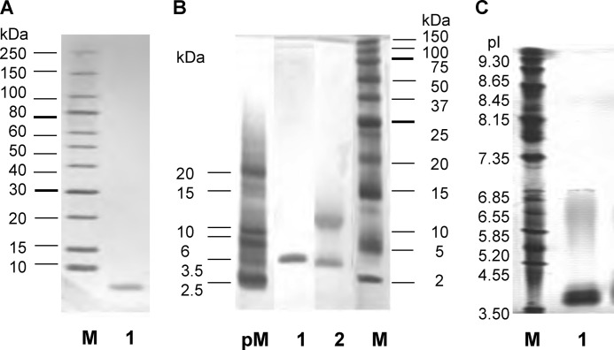
FIGURE 1.
Characterization of PhoSL. A, shown is an SDS-PAGE linear gradient gel (10–15%). Lane M indicates marker proteins; lane 1, purified PhoSL under reducing conditions with 2-mercaptoethanol. B, shown is a high density SDS-PAGE gel. Lane pM indicates marker peptides; lane 1, purified PhoSL under reducing conditions with 2-mercaptoethanol; lane 2, purified PhoSL non-reduced; lane M, marker proteins. C, shown is isoelectric focusing of PhoSL. Lane M indicates marker proteins; lane 1, PhoSL.

