FIGURE 3.
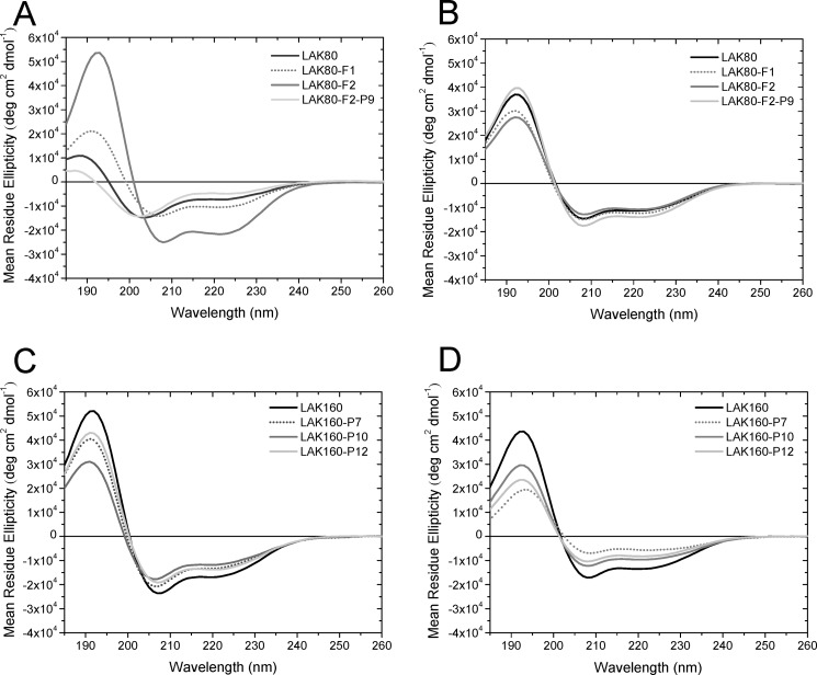
Circular dichroism spectra reveal the effect of peptide hydrophobicity and proline position on secondary structure in aqueous solution and membrane-mimicking medium. CD spectra are shown for LAK80, LAK80-F1, LAK80-F2, and LAK80-F2-P9 in 5 mm Tris buffer solution (A) or dimyristoyl phosphatidylcholine/dimyristoyl phosphatidylglycerol (75:25) liposomes (B) and LAK160, LAK160-P7, LAK160-P10, and LAK160-P12 in 50 mm SDS (C) or dimyristoyl phosphatidylcholine/dimyristoyl phosphatidylglycerol (75:25) liposomes (D). All spectra were recorded at 37 °C. deg, degrees.

