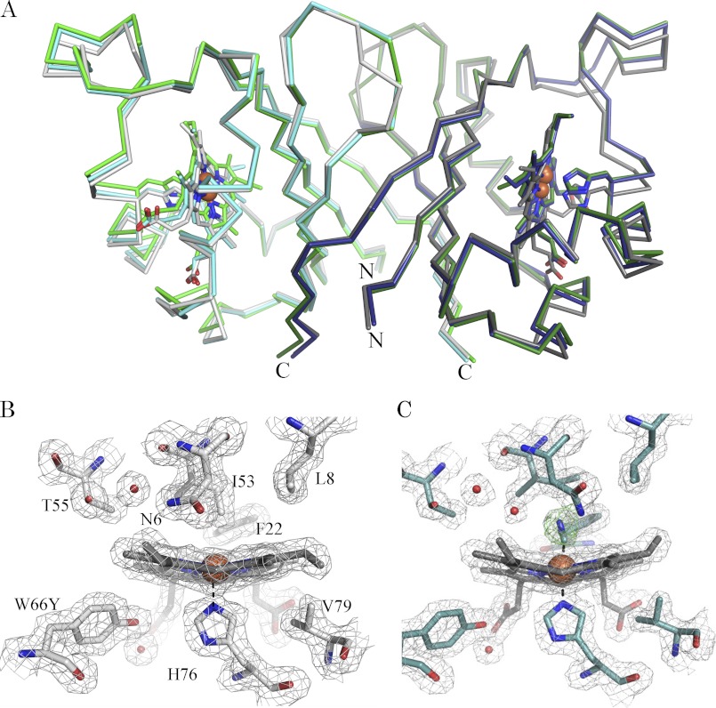
FIGURE 6.
Structural comparison of wild-type IsdI, the W66Y variant, and its cyanide derivative. A, the Cα trace of the structures of wild-type IsdI (PDBID 3LGN: green, chain A; dark green, chain B), IsdI-W66Y (gray, chain A; dark gray, chain B), and IsdI-W66Y(CN) (cyan, chain A; blue, chain B). The variant forms superimpose to wild type with root mean square deviations of 0.68 Å for IsdI-W66Y and 0.40 Å for IsdI-W66Y(CN). The N and C termini are shown. Shown are the 2 Fo − Fc map (gray) contoured at 1σ for the heme binding residues of chains A for the W66Y variant (gray) (B) and the cyanide derivative of the W66Y variant (cyan) (C). The omit map (green) for cyanide is contoured at 3σ.

