FIGURE 2.
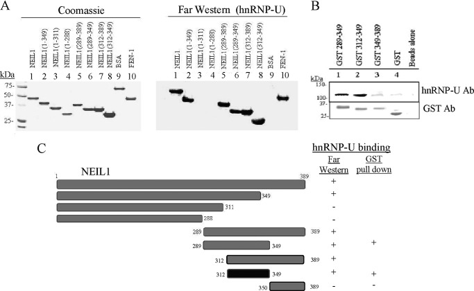
The hnRNP-U direct interaction with NEIL1 and identification of the NEIL1 C-terminal residues (312–349) as the minimal interaction peptide. A, far Western analysis. Left panel, membrane-bound WT NEIL1 (lane 1) and its deletion polypeptides (lanes 2–8; Coomassie staining after SDS-PAGE) were incubated with hnRNP-U in solution and probed with hnRNP-U Ab (right panel). BSA (lane 9) and FEN-1 (lane 10) were used as negative and positive controls respectively (22). B, co-elution of hnRNP-U with the NEIL1 GST-tagged deletion polypeptides bound to glutathione-Sepharose beads. C, the NEIL1 C-terminal residues (312–349) were identified as minimal interaction residues for hnRNP-U as shown schematically.

