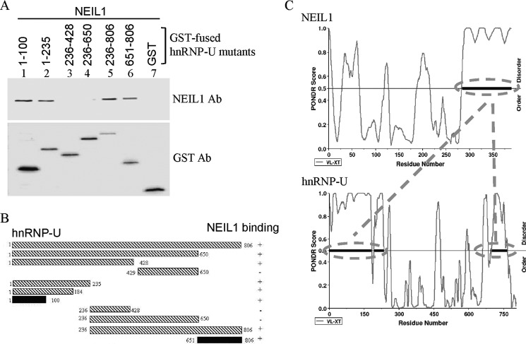
FIGURE 4.
Mapping and correlation of NEIL1 interaction domains in hnRNP-U with predicted disordered regions. A, co-elution of NEIL1 with GST-tagged hnRNP-U polypeptide fragments bound to glutathione-Sepharose beads and analysis of eluted proteins by Western analysis with NEIL1 Ab or GST Ab. B, schematic representation of the hnRNP-U N- (residues 1–100) and C- (residues 651–806) terminal segments for NEIL1 binding. C, PONDR modeling-based prediction of disordered structure of the interaction segments in NEIL1 (C terminus) and hnRNP-U (N and C termini), which was confirmed by CD spectroscopy (see supplemental Fig. S3).

