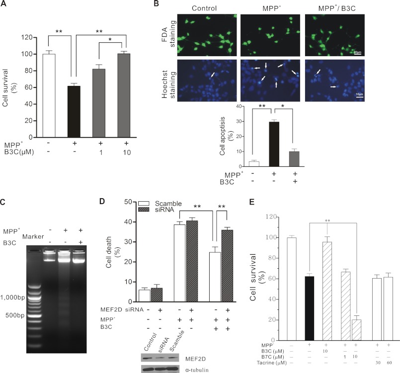
FIGURE 4.
B3C protects SN4741 cells from MPP+-induced apoptosis via MEF2D. A, attenuation of MPP+-induced death by B3C. SN4741 cells were preincubated with 1 or 10 μm B3C for 2 h and exposed to 500 μm MPP+ for another 24 h. Cell viability was measured using the MTT assay. Data were expressed as the percentage of untreated control (n = 3; *, p < 0.05, **, p < 0.01). B, SN4741 cells were treated as described in A. SN4741 cells were assayed with FDA and Hoechst 33324 staining. Viable cells were stained with fluorescein formed from FDA, which is de-esterified only by living cells. The number of apoptotic nuclei (white arrows) was counted and expressed as the percentage of total nuclei counted (n = 3; *, p < 0.05, **, p < 0.01). C, attenuation of MPP+-induced DNA fragmentation. SN4741 cells were treated as described in A. DNA was extracted from the SN4741 cells and analyzed by agarose gel electrophoresis and ethidium bromide staining. D, MEF2D-dependent attenuation of MPP+-induced death. SN4741 cells were transfected with scramble siRNA or MEF2D siRNA for 48 h and then treated as described in A. Cell viability was measured by lactate dehydrogenase releasing assay. Data were expressed as the percentage with total release after cell lyses set as 100% (n = 3; **, p < 0.01). The immunoblot shows the level of MEF2D after knockdown of MEF2D expression (Control indicates untransfected cells). E, B3C, but not B7C and tacrine, attenuates MPP+-induced SN4741 cell death. SN4741 cells were preincubated with B3C (10 μm), B7C (1 or 10 μm), or tacrine (30 or 60 μm) for 2 h and exposed to 500 μm MPP+ for another 24 h. Cell viability was measured using the MTT assay. Data were expressed as the percentage of untreated control (n = 3; **, p < 0.01).

