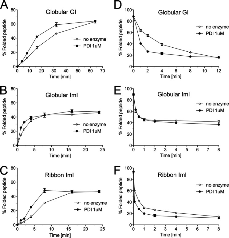
FIGURE 3.
Analysis of PDI-assisted folding and reduction of α-GI and α-ImI. A–C, oxidative folding was carried out in the presence of 0.1 mm GSH, 0.1 mm GSSG, and 20 μm linear peptide with and without Conus 1 μm PDI. D–F, peptide reduction studies were performed in the presence of 5 mm GSH, 20 μm folded peptide with and without Conus PDI. Folding and reduction reactions were terminated by acid quenching and analyzed by reversed-phase chromatography (C18 column, Vydac-Grace). Relative abundances of fully folded globular α-GI (A and D), globular α-ImI (B and E), and ribbon α-ImI (C and F) were determined from three independent experiments (mean ± S.D. (error bars)).

