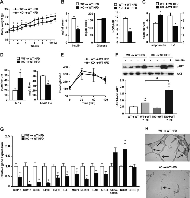
FIGURE 5.
Bone marrow-specific deletion of C/EBPβ reduced insulin levels and expression of adipose tissue inflammation and macrophage marker genes in HFD-fed mice. WT mice were transplanted with WT and C/EBPβ−/− bone marrow cells and their engraftment was tested at 10 weeks post-irradiation. At 4 weeks post-irradiation, mice were placed on HFD for 12 weeks. Data are mean ± S.E.; n = 6–7 mice/group. A, growth curve. B, fasting serum insulin (measured by ELISA) and blood glucose levels (measured by glucometer) and HOMA-IR. *, p < 0.05. C, serum levels of adiponectin and IL-6 were measured by ELISA and multiplex assay (respectively). *, p < 0.05. D, serum levels of IL-10 (measured by ELISA) and hepatic TG levels. *, p < 0.05. E, GTT. n = 3 mice/group. F, insulin-stimulated phosphorylation of Akt (Ser-473) in adipose tissue of BMT mice. Representative blots are shown. Graph under the blot shows quantification. Data are means ± S.E.; n = 4 mice/group. *, p < 0.05 versus control (non-insulin stimulated); #, p < 0.05 versus WT→WT + insulin. G, adipose tissue RNA was isolated, and relative gene expression was analyzed by qPCR. *, p < 0.05; n = 6–7 mice/group. H, adipose tissue samples (n = 3/group) from HFD-fed WT→WT and KO→WT animals were fixed in 4% formalin for immunohistochemistry as described under “Experimental Procedures.” Images were taken at ×200 magnification. Arrows point to crown-like rump structures within cells. Representative histology for both groups is shown.

