
FIGURE 3.
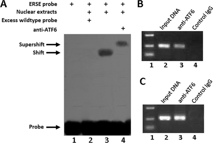
ATF6 binds to the promoter of the XBP1 gene. A, ATF6 binds to the XBP1 promoter in vitro (EMSA). Ten micrograms of nuclear extracts (NE) prepared from ATDC5 cells transfected with expression plasmid pcDNA3.1(−)-ATF6 were incubated with digoxigenin-labeled ATF6-binding site (ERSE) in reaction buffer (20 μl). For competition experiments, a 100-fold excess of wild type oligodeoxynucleotide was added. For supershift assays, anti-ATF6 (0.5 μg) was included. After 15 min of incubation, the digoxigenin-labeled probe was added, and the reaction mixture was incubated for an additional 15 min and analyzed by gel electrophoresis. The positions of the supershifted complex (supershift), the ATF6-ERSE complex (shift), and the free DNA probe (probe) are indicated. Arrows indicate free DNA probe (bottom) and DNA-protein complex (top). B, ATF6 binds to the XBP1S promoter in vivo (ChIP). C3H10T1/2 cells transfected with an expression plasmid encoding ATF6 were cross-linked by formaldehyde treatment and lysed. Cell lysates were subjected to immunoprecipitation with control IgG (lane 4) or anti-ATF6 antibodies (lane 3). Input DNA (lane 2; serving as positive control) and DNA recovered from the immunoprecipitation were amplified by PCR with the primers spanning the ATF6-binding site in the XBP1S promoter. C, ATF6 binds to the XBP1 promoter in the course of chondrogenesis. Micromass cultures of C3H10T1/2 cells were treated with 300 ng/ml BMP2 for 5 days, and cultures were processed and analyzed as in B.