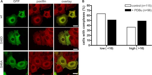
FIGURE 3.
GIT1 S46D localizes to paxillin-positive cytoplasmic complexes. A, MCF7 cells were plated onto collagen-coated coverslips and transiently transfected with expression vectors encoding GFP-GIT1 wt, S46A, and S46D as indicated. The next day, cells were fixed and stained with paxillin-specific primary antibody, followed by Alexa Fluor 546-conjugated secondary antibody (red). The images shown are projections of several confocal sections. Scale bar, 20 μm. B, MCF7 cells were plated onto collagen-coated coverslips and transiently transfected with an expression vector encoding GFP-GIT1 wt. Prior fixation, cells were left unstimulated or stimulated with 1 μm PDBu for 15 min. Cells were analyzed by confocal microscopy and scored according to their number of GIT1 cytoplasmic complexes.

