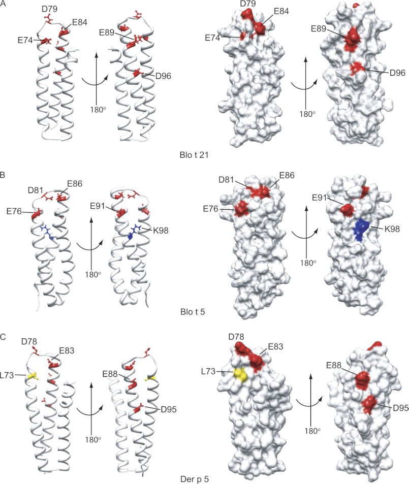
FIGURE 4.
Corresponding locations of the IgE-binding epitopes of Blo t 21 on the structures of Blo t 21, Blo t 5, and Der p 5. A, the mapped IgE-binding epitope residues of Blo t 21 are showed as ball-and-stick models on the ribbon diagram of Blo t 21, in two different orientations. The surface diagram of Blo t 21 shows that residues Glu-74, Asp-79, and Glu-84 form a surface cluster on one side of Blo t 21, whereas residues Glu-89 and Asp-96 form another cluster on the opposite side. B and C, the corresponding locations of the mapped IgE-binding epitope residues of Blo t 21 on the ribbon and surface diagrams of Blo t 5 and Der p 5, respectively. Positively charged, negatively charged, and hydrophobic residues are colored in blue, red, and yellow, respectively. Note that Asp-96 of Blo t 21 is replaced with Lys-98 in Blo t 5, whereas Glu-74 of Blo t 21 is replaced with Leu-73 in Der p 5. The figures were generated by the program Chimera (34).

