Figure 1.
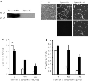
In vitro characterization of Synco-B18R. (a) Detection of B18R expression by far western blotting. Vero cells were infected with either Synco-B18R or Synco-2D for 24 hours before they were collected and lysed for detection of B18R by far western blotting as described in the Materials and Methods section. (b) Phenotypic characterization and comparison of Synco-B18R and Synco-2D. Vero cells were infected with Synco-2D or Synco-B18R at 0.1 pfu/cell and micrographs taken 24 hours later. Original magnification: ×200. UI, uninfected. (c,d) Virus replication in (c) SW480 and (d) HuH7 cells with or without IFN-α in the media. Cells were seeded in 24-well plates and infected with either Synco-2D (open bars) or Synco-B18R (closed bars) at 0.1 pfu/cell without or with IFN-α at the indicated concentration. Cells were harvested 24 hours later and the virus titer was determined by plaque assay. *P < 0.01 as compared with Synco-2D. There is no significant difference between Synco-B18R and Synco-2D in either of the cells in the wells without IFN-α in the media. IFN, interferon; pfu, plaque-forming unit.

