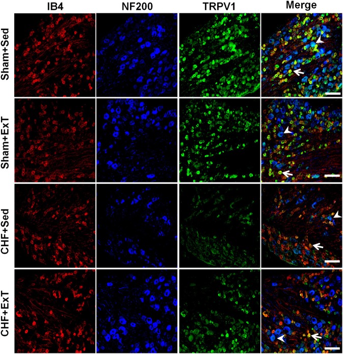
Figure 11.
Immunohistochemical data showing the protein expression of TRPV1 receptors in L4/L5 dorsal root ganglion (DRG) in sham and CHF rats. IB4, a C-fiber neuron marker; NF200, an A-fiber neuron marker. White Bar = 100 μm. White arrow represents double staining of TRPV1 with IB4, white arrowhead represents double staining of TRPV1 with NF200. [Reprinted from Wang et al. (2012). Copyright @ 2012 American Heart Association. Used with permission.]

