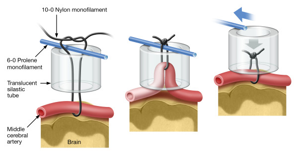
Figure 1.
Rat snare ligature model. The head of the rat was immobilized in a specially designed head holder. A skin incision was made between the eye and external auditory canal. The MCA was exposed by bisecting the temporalis muscle and performing a craniotomy anteriomedial to the juncture of the zygomatic arch with the squamosal bone. The dura was opened using a tuberculin syringe and the MCA was isolated from the arachnoid and pia by blunt dissection. Left Panel: construction of the snare ligature. Middle Panel: occlusion of the middle cerebral artery. Right Panel: removal of the snare ligature resulting in recanalization.

