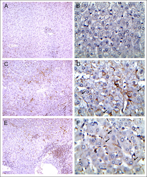
Figure 5.
Detection of galectin-9 in liver tissue sections.A, B) Normal liver tissue next to an adenoma (Table 2, patient #8, X100 and X400 respectively). Complete absence of galectin-9 staining. C, D) HCV-related liver cirrhosis next to a carcinoma (Table 2, patient #2, X100 and X400 respectively). Prominent staining is visible in Kupffer cells which are characterized by a flat triangle shape and their close association with sinusoid vessels. At high magnification, delicate, punctate staining is visible in a large number of hepatocytes (black arrows). E and F) HBV-related liver cirrhosis next to a carcinoma. (Table 2, patient #5) Predominant staining in various types of leucocytes often with a round shape. At high magnification, delicate punctate staining is visible in a large number of hepatocytes (black arrows).

