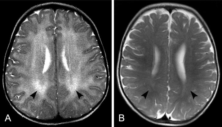
Fig 2.
Axial T2-weighted MRI scan of the brain of patient TTD421BE at age 19 months (A) compared to a normal MRI of a child at the age 13 months (B) using the same technique. A. The entire white matter of both cerebral hemispheres appears bright (arrowheads) compared to the darker cortical gray matter, indicating lack of normal myelin formation. B. In children with normal myelination, the signal intensity is reversed with the white matter being darker (arrowheads) than the gray matter in this type of scan. These images show that the myelin in the 13 month old child (B) is even more developed than that of our patient at 19 months (A).

