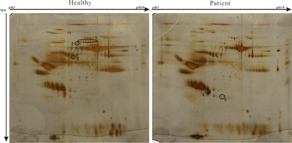
Figure 1.
2-DE gel patterns derived from healthy control and MG patient sera. Aliquots containing 100 μg of albumin/IgG-depleted proteins extracted from the sera of healthy controls (left) and MG patients (right) were subjected to isoelectric focusing (total: 38890 Vhs) using 13-cm strips (pH 3–10). The 2-DE gels were silver-stained. Differentially expressed proteins (> 1.5-fold) were labeled in the gels. The fold changes were calculated from three replicates. The X-axis shows pI 3–10 and the Y-axis shows the molecular weight in kDa.

