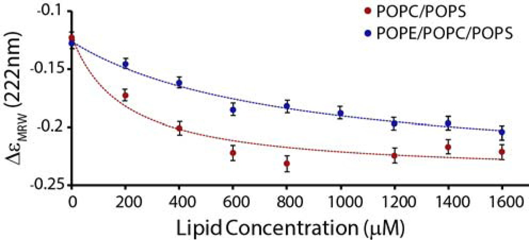
Figure 4.
Binding of prefibrillar IAPP to lipid vesicles. Changes in molar ellipicity at 222 nm arising from the coil-to-helix conformational change upon membrane binding as a function of the lipid concentration. 25 µM of freshly dissolved IAPP was titrated with the indicated concentrations of 7/3 POPC/POPS (filled circles) and 3/4/3 POPE/POPC/POPS (open circles) LUVs. The final conformation of IAPP is similar in both membranes (Fig. S1). Lines represent a binding isotherm to each dataset. Experiments were performed at 25 °C in 10 mM phosphate buffer, 100 mM NaF pH 7.4. Measurements represent the average of 30 measurements over 30 seconds, error bars indicate the standard deviation of this measurement.

