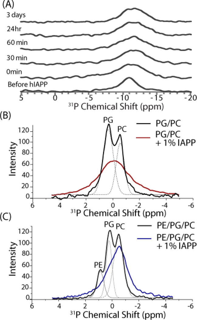
Figure 5.
31P NMR spectra revealing the interaction of IAPP amyloid fibers with lipid vesicles. (A) Time dependent static 31P NMR spectra of aligned bilayers after the addition of 1 mole % IAPP. Changes are not apparent in subsequent spectra after the addition of IAPP, suggesting IAPP reached the amyloid state before the first spectra was acquired. (B and C) Magic-angle spinning 31P NMR spectra of multilamellar vesicles (B) 7/3 POPC/POPG and (C) 3/4/3 POPE/POPG/POPC) incubated with 1.178 mM (1 mole %) IAPP. Dotted lines represent a deconvolution of the spectrum. Experiments were performed at 37 °C in 10 mM Tris Buffer, 100 mM NaCl, pH 7.4.

