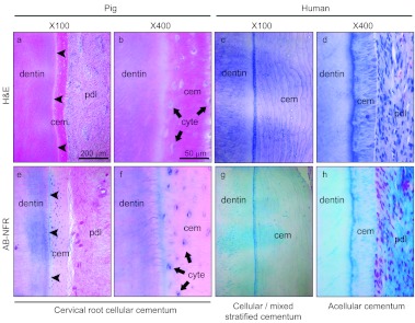
Figure 5.
Histological staining of porcine and human cementum. Sections of the first mandibular molar of a 13- to 16-week-old Hanford miniature pig and human third molar were used to contrast H&E and AB-NFR staining for visualizing cementum (CEM). (a and b) H&E and (e and f) AB-NFR staining of cellular cementum of the porcine cervical root provides contrast for observation of the CDJ (arrowheads in panels a and e) and cementum/PDL interface, as well as embedded cementocytes (cyte in panels b and f). In human molar, staining was evaluated for (c and d) mixed stratified cementum of the furcation area and (g and h) acellular cementum of the cervical root. Both H&E and AB-NFR provide contrast for the initial cementum layer in mixed stratified (c and g) and acellular cementum (d and h), while AB-NFR provides more contrast between acellular cementum and cells of the PDL. Original magnifications (×100 and ×400) are labeled over the panels. AB-NFR, Alcian blue and nuclear fast red; CDJ, cementum–dentin junction; H&E, hematoxylin and eosin; PDL, periodontal ligament.

