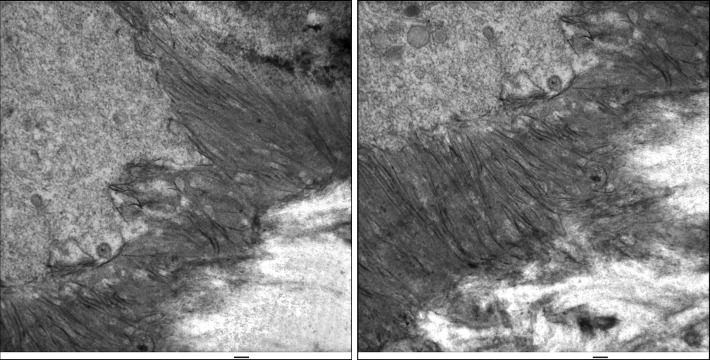
Figure 1.
Formation of initial enamel. Day 7 mouse mandibles were fixed with 2.5% glutaraldehyde in sodium cacodylate buffer and post-fixed with osmium tetroxide. Sections were stained with uranyl acetate, then lead citrate, and viewed by TEM. Ameloblasts are on the upper left. Banded collagen fibers are on the lower right. The enamel ribbons initiate on the dentin surface in close association with collagen and the mineralization front on the ameloblast membrane. Scale bars=100 nm. TEM, transmission electron microscopy.

