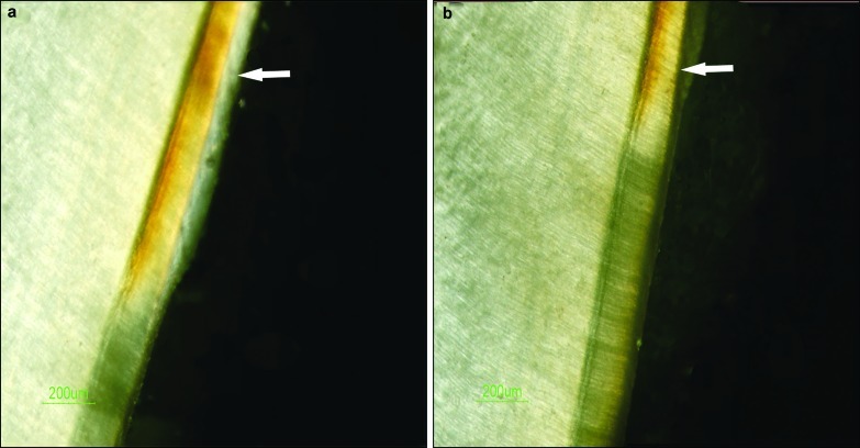
Figure 1.
The remineralization effect on erosive lesion observed by PLM. (a) Comparison of the erosive lesion before and after GCE remineralization. (b) Comparison of the erosive lesion before and after treatment with the remineralization solution. The arrows indicate the remineralization zones. The other transparent layers are the demineralization zones. PLM, polarized light microscopy; GCE, Galla chinensis extract.

