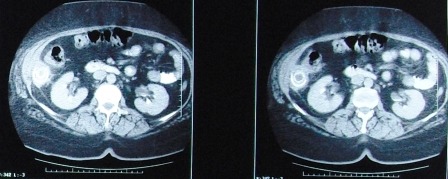
Fig. 1.

Abdominal CT scan shows a 50-60 mm, concentrically-layered, subhepatic collection with a dense, central, stone-like image, possibly sorrounded by the contrast material injected through the fistula; a segment VI hepatic abcess, exceeding the liver capsule, in direct contact with the walls of the right colonic angle and the ascending colon
