Figure 7. .
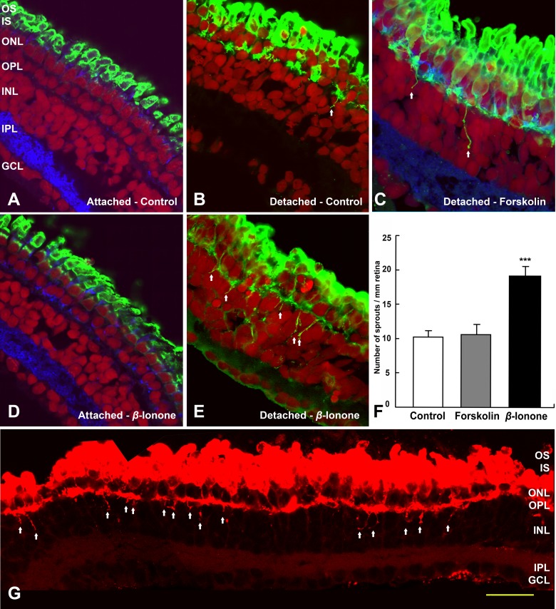
Rod cell sprouting in the detached but intact retina is increased by β-ionone after 7 days. (A) Salamander retina cultured in the eyecup show no rod opsin mislocalization and no rod cell sprouting. (B) Neural retina detached from the RPE show rod opsin mislocalization and some neuritic sprouting (arrows). (C) Detached neural retina cultured with FSK show rod opsin mislocalization and a little neuritic sprouting (arrows). (D) Retina cultured in the eyecup and treated with β-ionone show no rod opsin mislocalization and no sprouting. (E) Detached neural retina cultured with β-ionone show both rod opsin mislocalization and robust neuritic spouting (arrows). (A–E) Opsin, green; synaptophysin, blue; nuclei, red; ***P < 0.001). (F) Quantification of rod cell sprouting, from retinal sections immunolabeled for opsin after 7 days culture. The total number of sprouting neurites from a single retinal section was divided by total retinal length to yield a normalized number of sprouts. Treatment of detached retina with 10 μM FSK showed no significant increase in the number of rod cell sprouts compared with untreated, detached retina. In contrast, 10 μM β-ionone dramatically increased, by 87.2%, rod sprouts (n = 5 sections, 2 animals). (G) Detached but intact retina treated with 10 μM β-ionone. Images were reassembled into a complete retinal montage for each section; sprouts longer than 5 μm are indicated by arrows. Scale bar = 50 μm.

