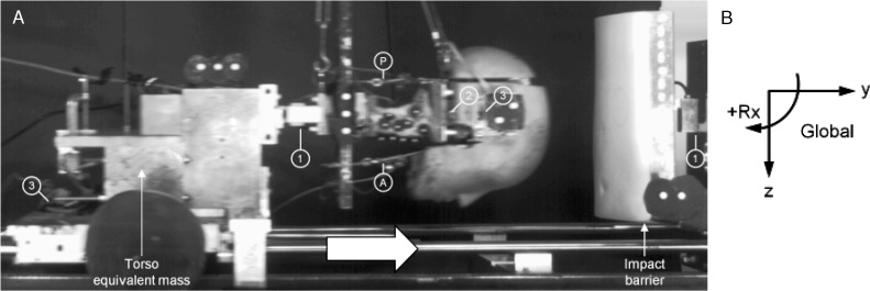
Figure 1.
A, High-speed camera photograph of the head and neck specimen during simulated head-first impact. The model consisted of a human cadaveric cervical spine specimen mounted to a mass on the sled representing the equivalent torso mass and carrying an anthropometric surrogate head. The head and neck were stabilized using anterior (A) and posterior (P) muscle-force replication cables. Motion-tracking markers were rigidly fixed to the head, cervical vertebrae, sled, and impact barrier. Load cells included 1 component between the base of the neck and torso mass and at the impact barrier (1) and 6 components in the head (2). Biaxial accelerometers (3) are rigidly fixed to the head and torso mass. B, The global coordinate system was fixed to the ground and had its positive z axis oriented anteriorly, positive y axis oriented superiorly, and positive x axis oriented to the left relative to the specimen. Sagittal rotation was positive for flexion and negative for extension.

