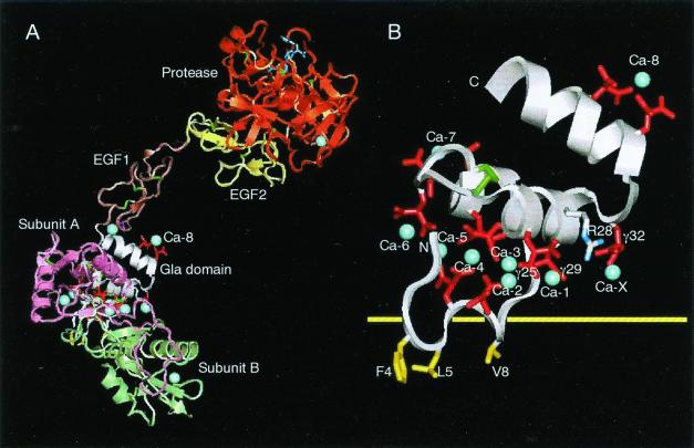
Figure 3.
Model of factor Xa bound to X-bp and XGD1-44 bound to membrane. (A) Factor Xa bound to X-bp. The Gla residues are in red, bound Ca2+ ions in blue (only labeled is Ca-8, which is identified in the present study), and disulfide bonds in green. The small molecule (dark blue) bound to the active site of the protease domain is the FX-2212a inhibitor (17). (B) Putative membrane-binding surface of XGD1-44. Same view as in A, but the scale of the figure is magnified for clarity. The hydrophobic patch includes Phe4, Leu5, and Val8, and hydrophilic patch includes Arg28, Gla25, Gla29, Gla32, and Ca-1 as a bridging Ca2+, which are on either side of the yellow horizontal line of the putative membrane surface. Ca-X is a putative Ca2+ ion that is taken in as another bridging Ca2+.

