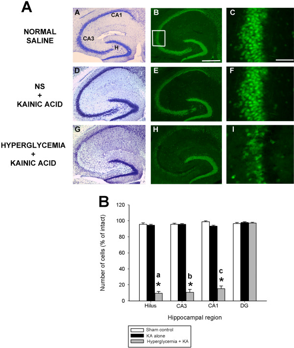
Figure 6.
Hyperglycemia aggravates status epilepticus-induced hippocampal damage. (A) Low-power photomicrographs of cresyl violet (A,D,G) and NeuN immunofluorescent-stained (B,E,H) horizontal sections of the hippocampus illustrating surviving cells throughout the hippocampus 7 days following systemic kainate administration to normoglycemic and non-ketotic hyperglycemic mice that underwent KA-induced SE. Hyperglycemia dramatically increased the extent of hippocampal cell death in excitotoxin cell death resistant B6 mice. Note the significant amount of cell loss, as evidenced by a loss of cresyl violet staining (G) and NeuN immunofluorescence (H), in representative sections from hyperglycemic KA-treated mice throughout all excitotoxin cell death susceptible regions (G,H,I) and absence of cell loss in KA-treated excitotoxin cell death resistant mice (D,E). CA1 and CA3 denote the hippocampal subfields; H, dentate hilus. Scale bar = 750 μm (A,C); 100 μm (B,D). High-magnification photomicrographs represent details of the boxed area of CA3 shown in B. (B) Quantitative analysis of neuronal density in hippocampal subfields following hyperglycemia + KA to B6 mice. Viable surviving neurons were estimated by cresyl violet staining. Bars denote the percentage of surviving neurons (as compared with saline-injected sham control B6 mice). Asterisks indicate significant difference compared with KA alone or Sham control of P<0.05. (ANOVA with post hoc Student Newman Keuls; a: F=781.34; P<0.001; b: F=542.49; P<0.001; c: F=567.89; P<0.001).

