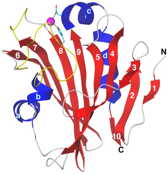
Figure 6. Modeled structure of spinach PsbP.

The structure is shown after 15 ns of molecular dynamics at 300 K. Secondary structure elements are indicated: strands, numbered blue; helices, lettered red; irregular, grey; modeled internal loops, yellow (longer loop, residues 90 to 107 between helix b and strand 6; shorter loop, residues 135 to 139 between strands 7 and 8). The Zn2+ ion (magenta sphere) is coordinated by Asp165 carboxylate and His144 imidazole (side chains in atomic colors with cyan carbons). The termini are labeled; the N-terminus is that of the crystal structure starting at residue 16.
