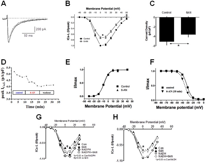
Figure 3. Inhibitory effects of 6AN on ICa-L in isolated rat cardiomyocytes and effect of NADPH on 6AN-induced inhibition of ICa-L.
A and B: ICa-L were evoked by 500-ms depolarizing pulses to 10 mV applied every 30 s from a holding potential of −80 mV. Cells were superfused with 6AN (5 mM) for 10 minutes after the currents had stabilized. Both the raw traces (A) and I–V relationships (B) show that ICa-L amplitudes were significantly reduced by 6AN. Currents were normalized to the peak amplitude at 0 mV in the absence of 6AN. Summary data for the normalized current densities (C) and time-course (D) indicate inhibition of ICa-L by 6AN. Stead-state activation (E) and inactivation (F) curves suggest 6AN had no significant effect on activation and inactivation kinetics. Summary data for the normalized current densities in cells dialyzed with NADPH (100 µM) and exposed to 6AN are shown. The I–V relationships indicate that ICa-L amplitudes were significantly reduced by 6AN (G), but that dialysis of NADPH partially reversed their inhibitory effect. In contrast NADH had no effect on 6AN-induced inhibition of ICa-L (H). Currents were normalized to the peak amplitude at 0 mV in the absence of 6AN. *P<0.05 vs. control. #P<0.05 vs 6AN.

