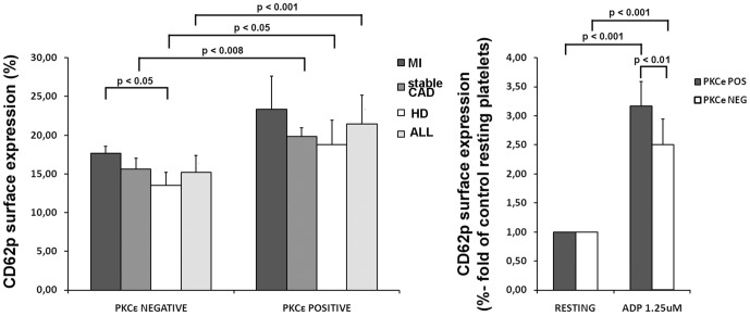
Figure 4. PKCε protein expression in platelets correlates with their activation levels.
Panel A: Flow cytometric analysis of platelet CD62p surface expression in patients with MI, sCAD, healthy donor and in all the sample (ALL), on the basis of PKCε expression. Cells were stained with specific mAb anti P-selectin (CD62p). Seven patients were analyzed for each group (MI: 2 PKCε negative and 5 PKCε positive samples; sCAD: 4 PKCε negative and 3 PKCε positive samples; HD: 4 PKCε negative and 3 PKCε positive samples). Data is expressed as mean ± S.D (Anova and Bonferroni t-test). Panel B: Flow cytometric analysis of CD62p surface expression in PKCε negative and positive platelets. Cells were treated with ADP and compared with untreated platelets (resting). Ten patients were analyzed for each group. Data is expressed as mean ± S.D (Anova and Bonferroni t-test).

