
Figure 2. Expression of MHC-I, IRF1, IRF2 and the NF-kB p65 subunit in primary NB lesions.
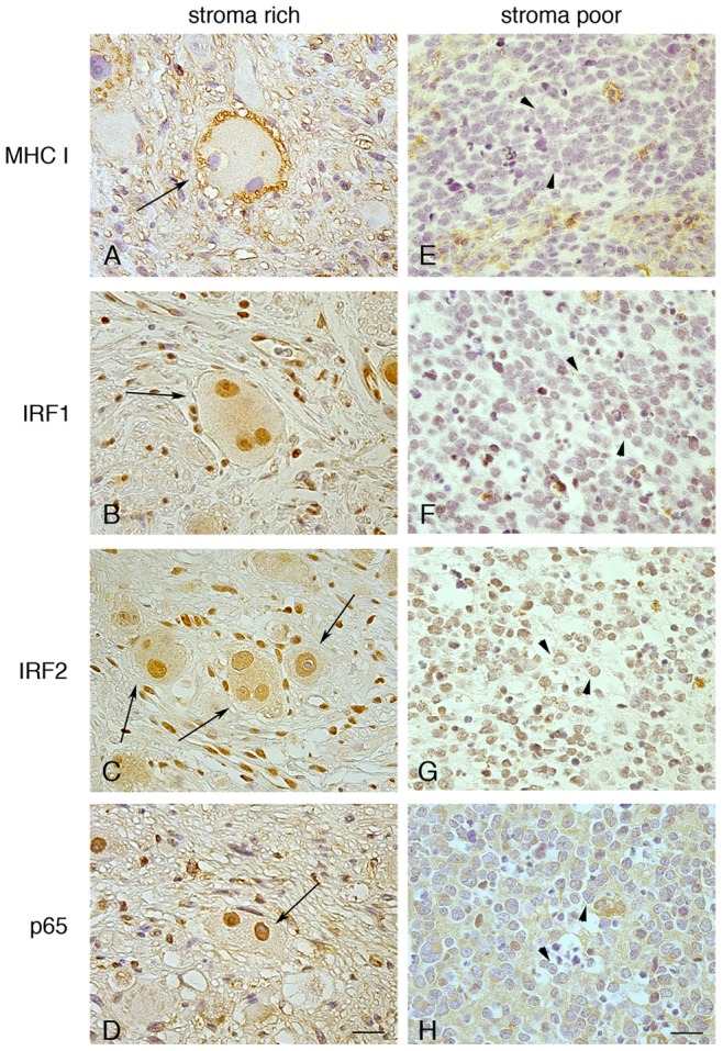
Immunohistochemistry of human NB tissue sections with Abs to MHC-I (A, E), IRF1 (B, F), IRF2 (C, G) or NF-kB p65 subunit (D, H). Visualized with diaminobenzidine (DAB; brown), nuclei counter-stained with haematoxilin (blue). IRF1, IRF2 and NF-kB p65 are strongly expressed in the nuclei of mature ganglion cells (arrows), endothelial cells, lymphocytes and stroma cells in the well-differentiated MHC-I-positive ganglioneuroblastoma (A-D), and weakly expressed in the MHC-I-negative neuroblastic cells (arrowhead), i.e. undifferentiated stroma-poor NB (E-H). In E-H, positive staining of benign cells, including lymphocytes and macrophages. NF-kB p65-positive staining of the fibrillary network in H is evident. Original magnification, x40. Scale bars 30 µm. Data shown are representative of 10 stroma-rich and 10 stroma-poor NB tissue sections.