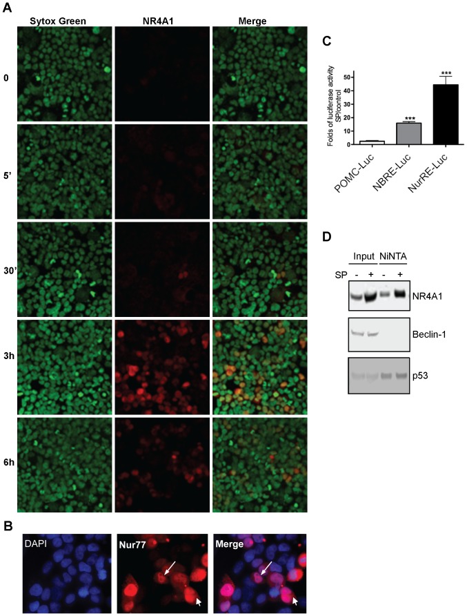
Figure 7. NR4A1 localized mainly in the nucleus during autophagic cell death, and is transcriptionaly competent.
HEK293 cells were transfected with NK1R and exposed to SP for the indicated time. A, Immunofluorescence of NR4A1 (red) showed it mainly in nuclei, which were stained with Sytox Green. 20× magnification. B, Examples of cells where NR4A1 (red) was found by immunofluorescence in both the cytoplasm and the nucleus (DAPI) after 3 hr of exposure to SP. 40× magnification. C, Luciferase essay to quantify NR4A1 transcriptional activity driven by either NBRE or NuRE containing promotor. Folds of luciferase activity after 6 hr of SP addition with respect to control cells are plotted. Bar represent standard deviation (n = 4, ***, p<0.001). D, NR4A1 interacts with p53 but not with Beclin 1. HEK293 cells were transfected with His-tagged NR4A1 and NK1R, and exposed or not to SP. NR4A1 was pulled down by NiNTA, and the presence of Beclin 1 or p53 complexed with NR4A1 was analized by Western blot.

