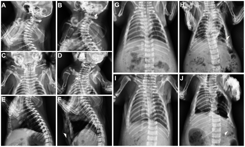
Figure 2. Malformations in the cervical and thoracic regions of VAD neonates.
(A) Side view of cervical region showing normal skeletal in a control fetus. (B) VAD fetus showing loss of the neural arch in cervical 1. (C) Ventral view of the scapula region showing normal development in a control fetus. (D) VAD fetus showing dysplasias of the scapula. (E) Side view of the sternal elements showing normal development the manubrium, sternebrae and xyphoid process in a control fetus. (F) VAD fetus showing malformation of the sternal elements as well as loss of xyphoid process. (G) Ventral view of the thoraric region showing normal development in a control fetus. (H) VAD fetus showing anomalies of vertebrae in thoraric region as well as rib fusions. (I) Ventral view of the thoraric region showing normal development in a control fetus. (J) VAD fetus showing loss of rib in vertebral 20.

