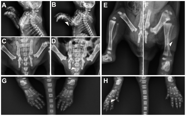
Figure 3. Malformations in the limbs, pelvic and sacral of VAD neonates.
(A) Side view of the forelimb region showing normal forelimb development in a control fetus. (B) VAD fetus showing deformities of ulna, at least unilaterally, in the forelimb region. (C) Ventral view of the pelvic region of a control fetus depicting normal development and attachment of the pelvic. (D) VAD fetus showing moderately dysplasia of the ischium. (E) Ventral view of the hindlimb region showing normal hindlimb development in a control fetus. (F) VAD fetus showing malformations of the tibia and fibula as well as tibia and fibula fusion. (G) Ventral view of the ossification region showing normal ossification development in a control fetus. (H) VAD fetus, the ossification of the second phalange was either missing or greatly reduced in size.

