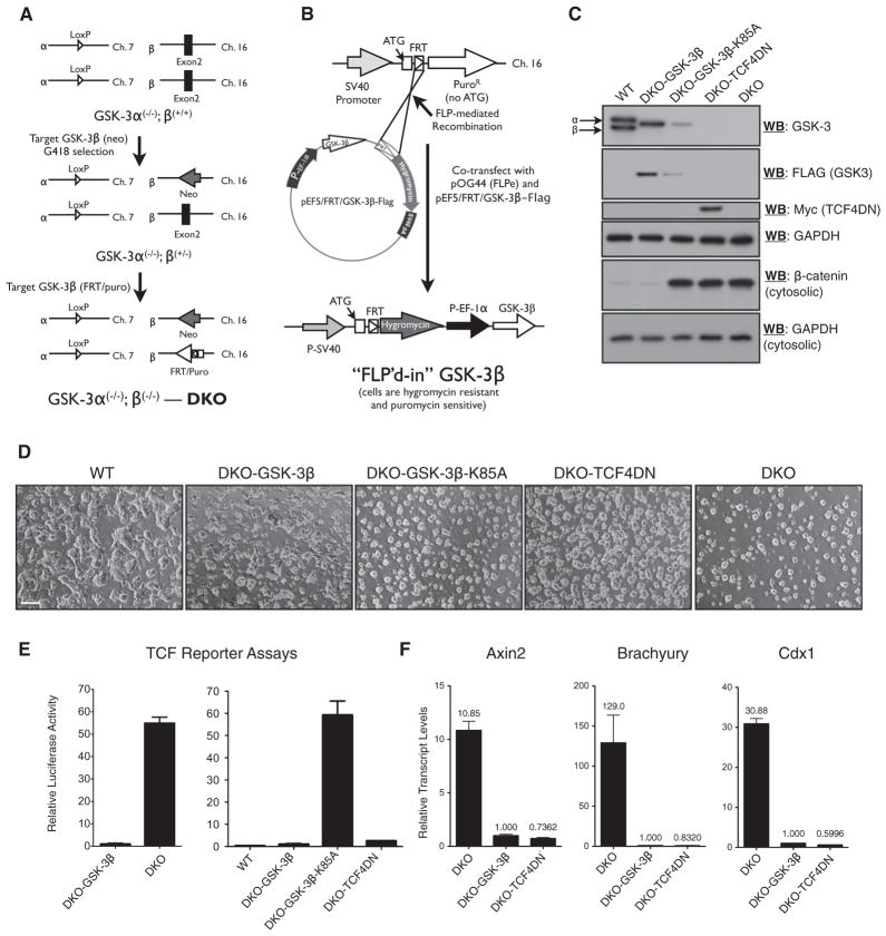
Figure 1. Generation and Validation of Isogenic DKO mESC Lines Expressing GSK-3 Variants or Dominant-Negative TCF4.
(A) Targeting steps used to create DKO mESCs, which incorporate an FRT recombination site for the Flp-in system in the GSK-3β locus.
(B) Flp-mediated site-specific integration of an EF-1α-driven transgene.
(C) Immunoblot analyses of transgene expression in mESC lines expressing TCF4DN or variants of GSK3.
(D) Morphology of WT, DKO, and DKO-Flp-in lines as indicated, via light microscopy. Scale bar represents 100 μm.
(E) TCF reporter assays demonstrate that re-expression of GSK3 in DKO mESCs restores steady-state inhibition of Wnt signaling (left) and that reporter activity is not altered by the expression of kinase-inactive GSK3 (K85A) but is efficiently attenuated by expression of TCF4DN in DKO cells (right).
(F) Quantitative RT-PCR analysis of Axin2, Brachyury, and Cdx1 transcript levels in the indicated cell lines.
Bars represent the mean of three independent experiments ± SEM. See also Figure S1.

