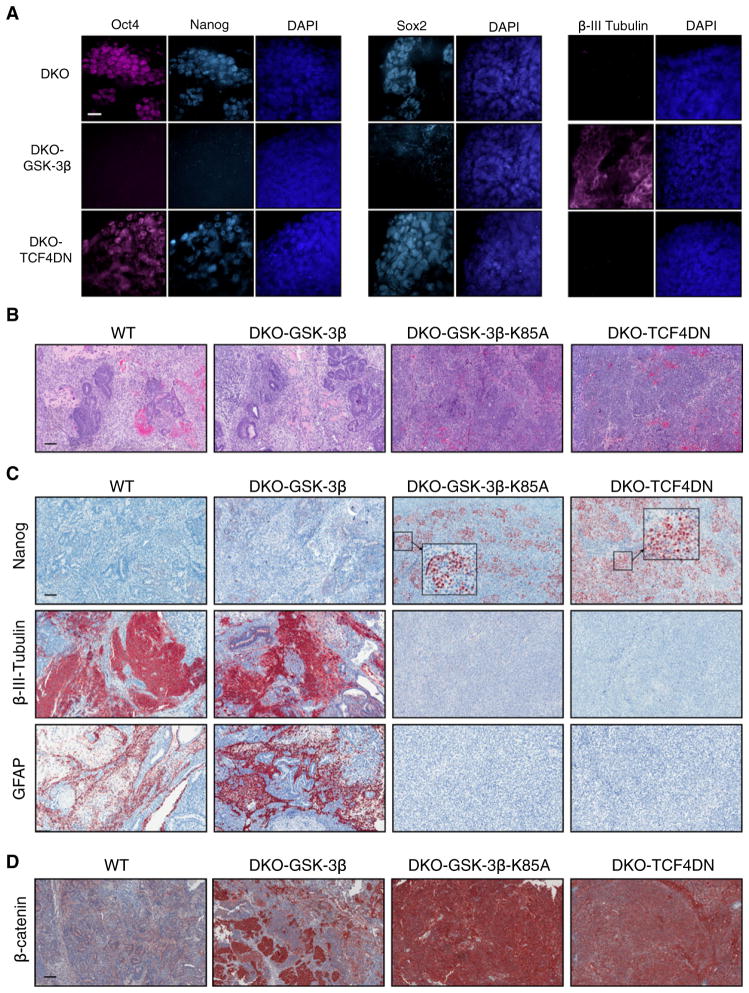
Figure 2. Attenuation of Wnt Target Gene Activation in DKO mESCs, by Stably Expressing TCF4DN, Fails to Rescue Their Impaired Differentiation.
(A) Retention of the pluripotency markers Oct4, Nanog, and Sox2 as well as expression of the neuronal marker β-III-tubulin was determined by immunofluorescent staining of the indicated EBs, maintained under differentiating conditions for 14 days.
(B) H&E staining of 3-week teratomas derived from WT (E14K), DKO-GSK3β, DKO-GSK3β-K85A, and DKO-TCF4DN mESCs.
(C) Immunohistochemical staining of teratomas revealed retention of the pluripotency marker Nanog and absence of neural tissue, as determined by staining for β-III-tubulin and GFAP, in teratomas derived from DKO-TCF4DN mESCs.
(D) β-catenin is maintained at low levels in teratomas derived from WT mESCs, whereas some regions of high β-catenin levels are observed in DKO-GSK3β teratomas, resulting from the loss of GSK3β transgene expression and reversion to DKO phenotype. In contrast, β-catenin levels remain high in DKO-GSK3β-K85A and DKO-TCF4DN teratomas.
Scale bars represent 20 μm (A) or 200 μm (B–D). See also Figure S2.

