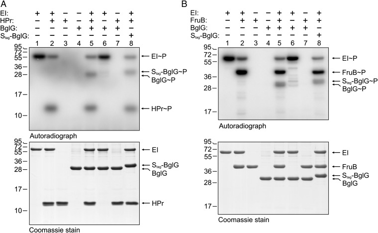
Fig. 3.
BglG becomes phosphorylated in vitro by HPr (A) as well as by FruB (B). PEP-dependent phosphorylation assays were carried out containing purified His10–EI, His10–HPr, Strep–BglG, or Strep–Stag–BglG as indicated. In the assays shown in B, His10–HPr was replaced by His10–FruB. Assays were carried out using either [32P]-PEP (Upper) or 1 μM cold PEP (Lower). Proteins were subsequently separated by denaturing gel electrophoresis using 15% (A) or 12% polyacrylamide gels (B). Gels were analyzed by phosphoimaging (Upper) or staining with Coomassie brilliant blue (Lower). Positions of the molecular weight marker are given at Left.

