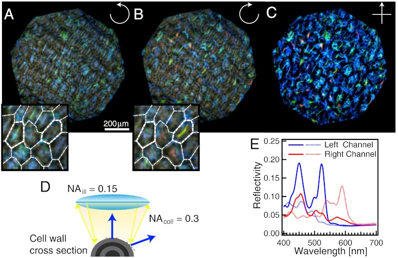
Fig. 3.
Polarized reflection of Pollia condensata fruit. (A) LH and (B) RH optical micrographs of the same area of the fruit under epi-illumination. The insets show a zoom of the central areas, with white lines delimiting the cells. (C) The same area of the fruit surface was also imaged between crossed polarizers. All three images were obtained using a × 10 objective. (D) Schematic representation of light reflection from a curved multilayer, representing the ellipsoidal shape of the epicarp cells. Only light from the central part of the cell is reflected into the numerical aperture of the objective (NA = 0.3), resulting in a color stripe in the center of each cell, seen in A and B. (E) Spectra from two different cells (continuous and dotted lines, respectively) for the two polarization channels (red and blue color, respectively). Auxiliary minor spectral features leading to the double-peak structure arise from the stacked nature of the cells in the epicarp (Fig. 2B). This leads to spectral contributions from underlying cells with different p values.

