Fig. P1.
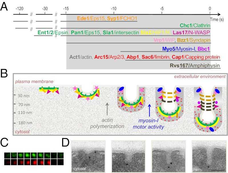
(A) Sequential recruitment of proteins (yeast/mammalian homologs) to the endocytic sites as assessed by live-cell fluorescence microscopy (4). The color indicates groups of proteins with similar distributions along the endocytic invaginations in our EM study, which is summarized in B. Steps block by drugs preventing actin polymerization or mutations in the myosin-I ATPase are indicated. (C) Fluorescence micrographs of consecutive 300 × 300-nm frames from double color time-lapse movies showing the transient recruitment of fluorescent GFP-Myo5 (green) and Abp1-RFP (red) at cortical endocytic in living cells. Frames were recorded every 2 s. Modified from ref. 5. (D) Electron micrographs of plasma membrane invaginations of increasing length specifically labeled with immunogolds (black dots) against endocytic proteins on ultrathin sections of chemically fixed yeast. (Scale bar: 100 nm.)

