Fig. P1.
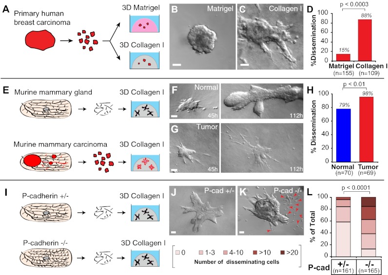
The ECM regulates the dissemination of mammary epithelial cells. (A) Schematic of the isolation and 3D culture of human breast tumor organoids. (B and C) Representative images of human tumor fragments in (B) Matrigel and (C) collagen I. (D) Percentage of tumor organoids showing cell dissemination in Matrigel and collagen I. (E) Schematic of isolation and 3D culture of normal and tumor murine mammary organoids in collagen I. (F and G) Representative time-lapse images of (F) normal and (G) tumor organoids in collagen I. (H) Percentage of normal and tumor organoids that disseminated cells into collagen I. (I) Schematic of organoid isolation from P-cadherin+/− and P-cadherin−/− mouse mammary glands. (J and K) Representative images of (J) P-cadherin+/− and (K) P-cadherin−/− organoids in collagen I. (L) Distribution of the number of disseminated cells per organoid in P-cadherin+/− and P-cadherin−/− epithelia. n = total number of organoids. (Scale bars, 50 μm.)

