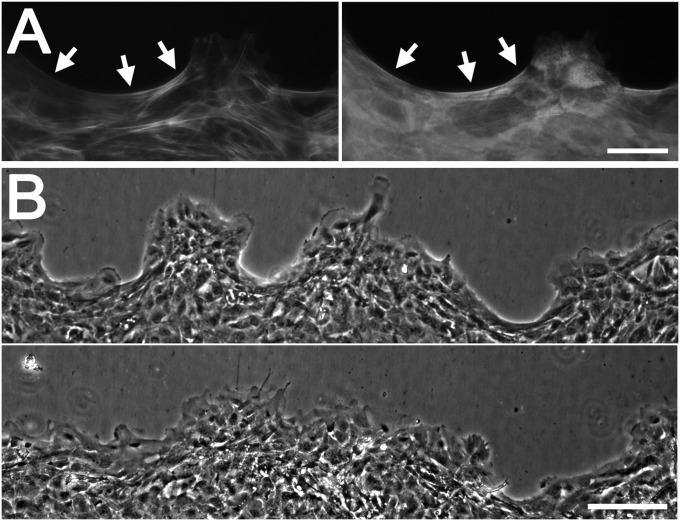
Fig. 3.
Role for purse strings in movement of epithelial sheets. (A) Cells at a moving epithelial edge were stained with labeled phalloidin (Left) or antibodies to myosin IIA (Right). Arrows indicate purse-string–like structures at concave regions. (B) Edge at the time of addition of 0.25 μM tyrphostin AG 1478 (Upper) and the same region 14 h later (Lower). (Scale bars: 100 μm.)

