Fig. 4.
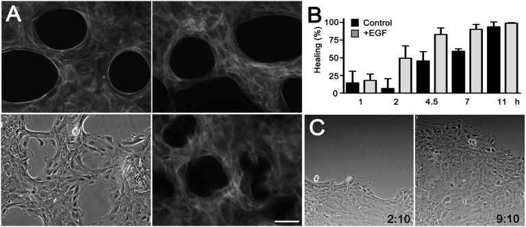
Switch to the crawling mode of healing by EGF signaling. (A) Cells were grown around agarose droplets and were either untreated (Upper Left) or treated with 10 nM EGF for 10 h without removing the agarose droplets (Upper Right) and stained with Alexa Fluor 546–conjugated phalloidin. Cells were incubated with 10 nM EGF and photographed in phase contrast (Lower Left) or stained with phalloidin 3 h after removal of agarose (Lower Right). (Scale bar: 100 μm.) (B) Healing of holes after removal of agarose droplets. EGF (10 nM) was added where indicated. Means of triplicates ± SD. (C) Progression of moving epithelial sheets at the time of addition of 10 nM EGF and 7 h later (Movie S7).

