Fig. 2.
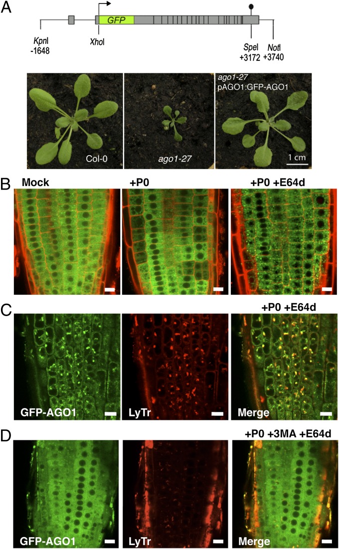
Subcellular localization of AGO1 along its degradation process. (A) The pAGO1:GFP-AGO1 construct complements ago1-27 allele phenotype. (B) Subcellular localization of functional GFP-AGO1 assayed by confocal microscopy. Seven-day-old seedlings were transferred from MS-agar plates to liquid MS medium supplemented with the indicated drugs and observed after overnight incubation (16–18 h). In the root tip of XVE-P0BW/GFP-AGO1 reporter lines GFP-AGO1 is localized exclusively in the cytoplasm of cells (Left). After P0 induction, the GFP-AGO1 signal decreases with a nonhomogenous pattern from cell to cell and is relocalized in vesicular-shaped structures (Middle). When P0 induction is combined with E64d (20 μM) treatment, GFP-AGO1 is stabilized and massively accumulates in these vesicular-shaped structures (Right). These speckles colocalize with acidic vesicles labeled with LysoTracker Red DND-99 (LyTr) (C) and their formation is significantly reduced if P0 induction and E64d treatment are combined with 3-MA (5 mM) (D). (Scale bars: 10 μm.)

