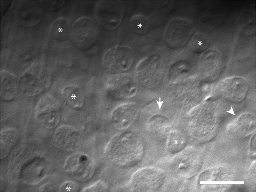
Figure 1.
Microscopic targeting of transient ON-OFF RGCs. Gradient-contrast optics micrograph of the RGC layer in the visual streak of the isolated rabbit retina. The arrow marks a small, round soma that is typical of transient ON-OFF RGCs. Displaced starburst amacrine cells are also numerous in the RGC layer but have slightly smaller somas that are almost filled by the nucleus (asterisks). The arrowhead marks a more elongated soma that is typical of a local edge detector RGC. Scale bar = 20 µm.

