Figure 5.
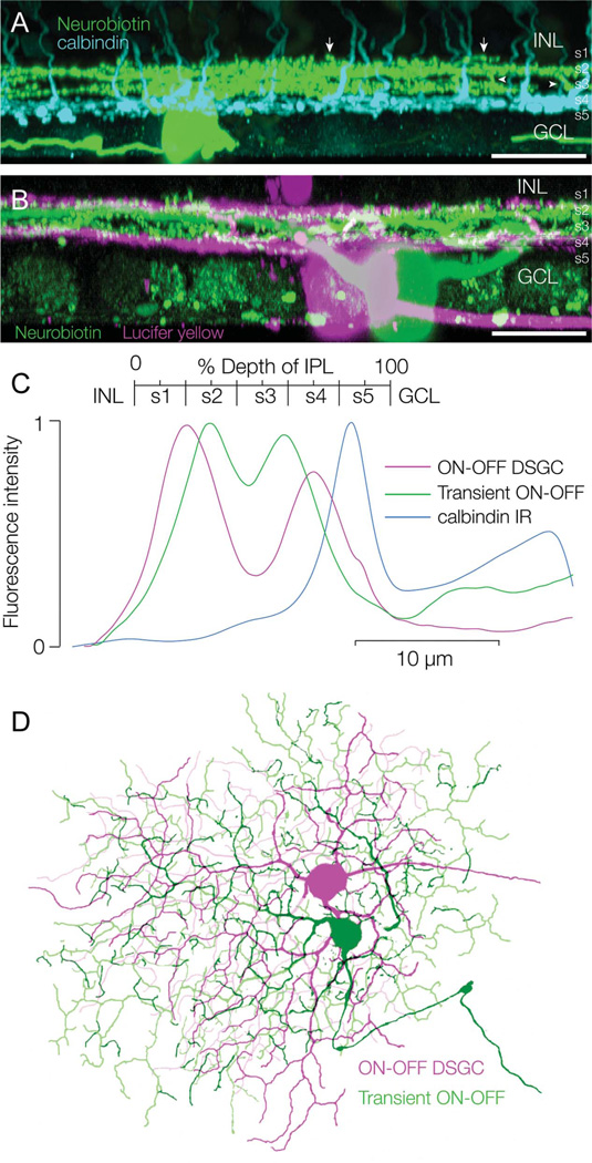
Stratification of transient ON-OFF RGCs. A: Side projection of a transient ON-OFF RGC labeled with Neurobiotin (green) following physiological identification. The retina is double labeled with an antibody against calbindin, which labels a population of bipolar cells (cyan). The RGC dendrites are bistratified and located above the axon terminals of the calbindin bipolar cells, which branch around the S4/S5 border. The ON and OFF arbors in S3 and S2 interconnect through vertical branches (arrowheads) and some OFF dendrites branch in S1, above the main OFF arbor (arrows). B: Side projection of a transient ON-OFF RGC labeled with Neurobiotin (green) and an overlapping ON-OFF DSGC labeled with Lucifer yellow (magenta); the arbors of the transient ON-OFF RGC stratify between the arbors of the ON-OFF DSGC. C: Mean fluorescence taken from z-sections through the ON-OFF DSGC (magenta) and transient ON-OFF RGC (green) in B and though the calbindin bipolar cells in A. D: Confocal reconstruction of overlapping dye-filled RGCs shows that the dendritic tree of the transient ON-OFF RGC (green) is slightly larger than that of the ON-OFF DSGC (magenta). Scale bars = 50 µm.

