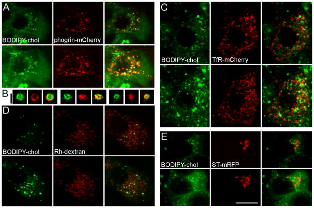
Figure 1.
BODIPY-chol is localized to insulin granules in live β-cells. MIN6 cells were transfected with phogrin-mCherry to label insulin granules (A and B, red), TfR-mCherry to label the recycling endosomes (C, red), ST-mRFP to label the TGN (E, red), or incubated overnight with rhodamine (Rh)-dextran to label the lysosomes (D, red). Cells were then labeled with BODIPY-chol (green in all panels) for 3 h at 37 °C and imaged live by confocal microscopy. The top row in each panel shows individual confocal planes; the bottom row shows projections of all confocal planes in a z-stack. (B) Three examples of super-resolution SIM images of phogrin-mCherry and BODIPY-chol localized to the same granule. The white scale bar, 10 μm, applies to A, C, D, E; Black scale bar for B: 0.5 μm.

