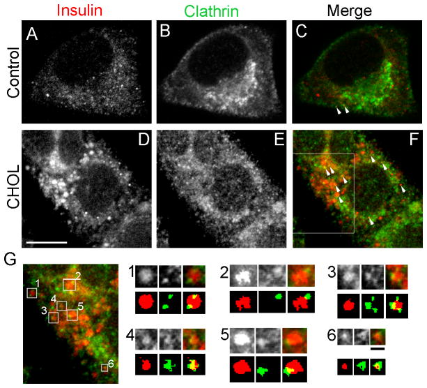
Figure 8.
More clathrin is associated with cholesterol-overloaded insulin granules. Control and cholesterol-overloaded (“CHOL”) MIN6 cells were immunostained with insulin (A and D, red) and clathrin (B and E, green) antibodies. (C, F) Structures positive for both insulin and clathrin are highlighted by white arrowheads. White bar, 5 μm. (G) A region of the cell marked by the white box in (F). The top row of the panels in (G) shows enlarged images of insulin granules displaying regions overlapping with clathrin. The numbers correspond to the squares in (G). The bottom row outlines the relevant structures labeled by each antibody, with background fluorescence removed by thresholding. Black bar, 0.5 μm.

